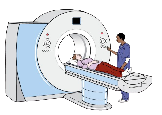
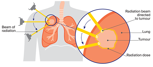
Radiation Basics

Leaf Decoration Leaf Decoration

What Type of Radiation is Used in Diagnosis and Treatment?

Diagnosis: CT

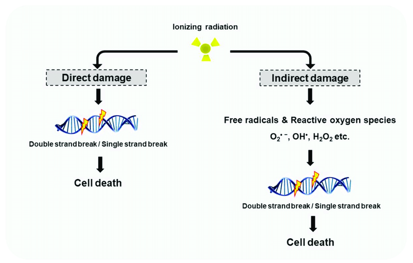
CT uses x-rays, which is a form of ionizing radiation. Ionizing radiation is defined as radiation that has undergone ionization (Hall & Giaccia, 2019).

  • Ionization occurs when radiation has enough energy to eject an orbital electron from a molecule or atom.
  • Large amounts of energy being released locally is a key characteristic of ionizing radiation (Hall & Giaccia, 2019).
CT Scan

Treatment: Radiation Therapy

External beam radiation therapy uses a radiation beam with photons (x-rays), protons, and electrons (National Cancer Institute, 2018).

  • Photons
    • High penetrating ability, allowing beam to reach tumours deep in the body
    • Travel into the patient’s tissue after hitting a tumour
    • Much higher energy levels relative to photons in x-ray imaging
    • Undergo scatter along their path
  • Protons
    • Particles with a positive charge
    • High penetrating ability, allowing beam to reach tumours deep in the body
    • Beam stops after hitting a tumour, reducing dose to surrounding healthy tissue
    • Does not undergo scatter along its path
  • Electrons
    • Particles with a negative charge
    • Limited to superficial tumours and skin lesions due to low penetrating ability
Radiation Therapy

How Does Ionizing Radiation Interact with Different Cell Types and Critical Structures?

  • No visible signs or symptoms for doses within the diagnostic imaging range.
  • Signs and symptoms from doses associated with radiation therapy include: fatigue, difficulty breathing, nausea, vomiting, skin irritation, scarring of lung tissue, and more (Lee, 2020).
  • Radiation Therapy
  • X-rays are sparsely ionizing (low LET), undergoing indirect action (Hall & Giaccia, 2019).
    • Indirect action occurs when radiation interacts with other atoms or molecules (e.g., water), creating free radicals that can diffuse and damage critical targets.
  • Ionizing radiation causes DNA single-strand breaks (SSB) and double-strand breaks (DSB) (Devic et al., 2022).
    • Free electrons either directly interact with cellular DNA or form free radicals, causing SSBs or DSBs (Huppmann et al., 2010).
    • If the cell cannot repair these breaks, loss of function or cell death may occur. Mutations can lead to genetic (germ cell) or somatic (cancer) effects (Huppmann et al., 2010).

Risks

CT

  • CT has a large effective dose of about 15 mSv for a chest CT scan. Since larger volumes of tissue are exposed than during a standard x-ray, the dose is higher than for a chest x-ray. However, the better contrast resolution and detail often outweigh the risks (Hall & Giaccia, 2019).
  • The most extensive cause of medical exposure to ionizing radiation is CT (Devic et al., 2022).
  • From the low doses associated with diagnostic imaging, risk estimates for mortality and radiation-induced cancer are based on Hiroshima/Nagasaki survivor data. The BEIR VII Committee states there is a linear, no-threshold dose–response for ionizing radiation and cancer (Huppmann et al., 2010). This means that any amount of radiation may result in cancer formation.
  • Developing cancer from any cause already has a probability of 42/100. This high background incidence makes it difficult to precisely quantify excess risk from diagnostic and therapeutic levels of ionizing radiation (Huppmann et al., 2010).
  • Although breasts and lungs are the most likely targets of radiation-induced cancers in adults, the thyroid absorbs the most dose. However, the thyroid has lower cancer risk due to being more radioresistant as it shrinks with age (Huppmann et al., 2010).
  • Radiation doses from most diagnostic imaging are too low to cause deterministic effects. However, genetic mutations can lead to stochastic effects (e.g., cancer) with no minimum threshold. Probability of occurrence increases with dose (Huppmann et al., 2010).
  • The overall five-year survival rate for lung cancer is ~16%. Early-stage cases are often detected incidentally. Thus, implementing CT scans for lung cancer screening is very beneficial (Huppmann et al., 2010).
  • Chest CTs for , instead of diagnosis use relatively lower doses (~0.65 mSv) (Huppmann et al., 2010).
  • CT Scan

    Image Credit: dialitdown.org

  • The International Commission on Radiological Protection estimates that 3 of every 100,000 screened men and 6 of every 100,000 screened women may develop radiation-induced cancer over 15–20 years (Huppmann et al., 2010).
  • Contrast-enhanced CT may cause rare allergic reactions (itching, hives, SOB, throat swelling). Contrast can also affect kidneys in those with preexisting impairment. Precautions include checking kidney function (blood test) and allergy history before administering (National Cancer Institute, 2024).

Radiation Therapy

  • During radiation therapy, the healthcare team protects healthy cells in the treatment area as much as possible. However, some damage to healthy cells may still occur, causing side effects. These can appear during treatment, right after, or weeks later. Most resolve or can be managed, but some can persist or become permanent (Lee, 2020).
  • Radiation therapy induces DNA breaks, leading to cell death (Majeed & Gupta, 2023).
    • Cancer cells are more severely affected than non-cancer cells.
  • Free radicals from ionizing radiation damage cellular components, leading to chromosomal aberrations and DSBs (Majeed & Gupta, 2023).
    • These changes cause cells to become lethally damaged. They may continue to divide before dying via mitotic cell death or apoptosis.
    • Mitotic cell death occurs due to improperly repaired chromosomal damage; apoptosis is due to direct damage.
    • The latent period before acute side effects is due to this delay in cell death.
    • Fibrosis and scarring can occur if stem cells differentiate into fibroblasts as a response to irradiation.
    CT Scan

Comparison of Radiation Exposure to Other Events

  • One chest CT scan for diagnosing SCLC is ~1.6 mSv (males) or 2.1 mSv (females). Females get a higher dose due to breast tissue (Larke et al., 2011).
  • Although cancer formation is a stochastic effect with no threshold, the dose from medical imaging is still lower than the ~3 mSv average yearly background dose (Lin, 2010).
  • The NCCN recommends radiation therapy for lung cancer at either 45 Gy total (1.5 Gy fractions, BID x3 weeks) or 60–70 Gy total (2.0 Gy fractions, QD x6–7 weeks) (Farrell et al., 2019). Fractionation allows cells time to repair.
  • Natural background radiation:
    • Average annual cosmic ray dose ~0.26 mSv at sea level, doubling every 2000 m altitude (Hall & Giaccia, 2019).
    • Earth’s crust dose ranges 0.15–1.40 mGy/year, region/altitude dependent (Hall & Giaccia, 2019).
    • Potassium-40 in some foods (e.g. bananas) ~0.2 mSv/year (Hall & Giaccia, 2019).
    • Radon is the largest source of natural background radiation, causing ~10% of all lung cancer deaths (15k–21k people) (Hall & Giaccia, 2019).
  • Chernobyl (1986):
    • Evacuated residents near the plant had ~30 mSv average effective dose (Government of Canada, 2022).
    • In Belarus, Russia, Ukraine: ~9 mSv average effective dose to the general public (Government of Canada, 2022).
    • Recovery workers on site 1986–1990 had doses averaging ~120 mSv (Government of Canada, 2022).
  • Radiation Compare 2

    Image Credit: London Cardio Clinic

References