There are many methods our radiographic procedures ensure radiation safety for the patients.
- Use of Lead
- We provide both lead aprons and lead collars when appropriate during radiographic exams.
- The lead, around 0.5mm thick, is capable of reducing scatter radiation by about 90% (Cheon et al., 2018).
- Radiographic procedures themselves are rather low doses of radiation (range), so effects are already unlikely (Hall & Giaccia, 2019).
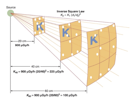
- Distance
- Inverse-square law (Lakhwani et al., 2018)
- The intensity of the X-ray beam is proportional to the reciprocal of the distance squared.
- The further away someone is from the radiation source, the lower the intensity of radiation they receive.
- Radiation technologists try their best to optimize distance so there is sufficient exposure for good image quality while minimizing dose to patients.
- Inverse-square law (Lakhwani et al., 2018)
- Fractionation
- Primarily used in radiation therapy rather than radiography.
- Fractionating dose delivery allows for cells to recover while the area of interest still receives a sufficient dose for treatment (Hall & Giaccia, 2019).
- This limits the effects on surrounding tissues, while effectively targeting the treatment area.
- ALARA Principle
- This is the “As Low As Reasonably Achievable” principle, where hospitals and radiation workers work to limit exposure to patients as much as possible without compromising image quality.
- A study conducted by Safrullah et al. (2017) concluded that around 99% of hospitals that completed their survey recognized the ALARA principle.
- 84% affirmed the beneficial use of lead protection (Safrullah et al., 2017).